
Early attempts in the surgical correction of refractive errors trace back to the mid-late nineteenth century and were employed in folk medicine and referenced in the European scientific literature. Of particular note is the work of Dr. Lans from the Netherlands in the 1890s. Further advances can be traced back to the 1930′s in Japan and 1970s, in the Soviet Union. Development of a variety of procedures such as Radial Keratotomy, Keratomileusis, Thermokeratoplasty, and Intraocular lens refractive surgery continued over the following decades. In the late 1970s scientists at IBM developed an ultraviolet laser to etch computer microchips. In 1983, Dr. Trokel at Columbia University and Dr. Srnivasan from IBM first introduced excimer laser corneal surgery. Dr Pallikaris first reported LASIK in 1990. Currently employed techniques include LASIK, PRK, Astigmatic Keratotomy and Refractive Lensectomy.
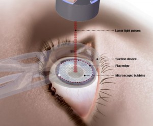
LASIK stands for “Laser Assisted In Situ Keratomileusis.” This literally means “to shape the cornea within, using laser” LASIK can be used to reduce nearsightedness (myopia), farsightedness (hyperopia) or astigmatism.
For example: In nearsighted people, the eye is too large, causing images to focus in front of the retina.
This steep curvature causes blurred images in the distance. LASIK removes microscopic layers in the cornea. Near sighted LASIK removes microscopic layers in the cornea resulting in a “flatter” corneal surface. Images can fall directly on the retina in the back of the eye to create improved, natural vision.
In farsighted people the eye is too small causing images to focus behind the retina. This causes blurred images up close and as we age, at a distance. This is not to be confused with presbyopia. (Presbyopia is what happens to all of us after we reach the age of 40… But more on that later.) Farsighted LASIK removes microscopic layers in the outer part of the cornea resulting in a “steeper” corneal surface.
In patients with astigmatism, the cornea has a toric shaped surface similar to that of a football. If you look at a football, there are 2 radii of curvature 90 degrees apart. When a patient with astigmatism looks at an object, this object is “split” into 2 images. Patients with high degrees of astigmatism wear hard contact lenses or glasses. Lasik correction for astigmatism removes the astigmatism.